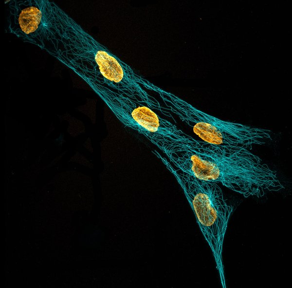
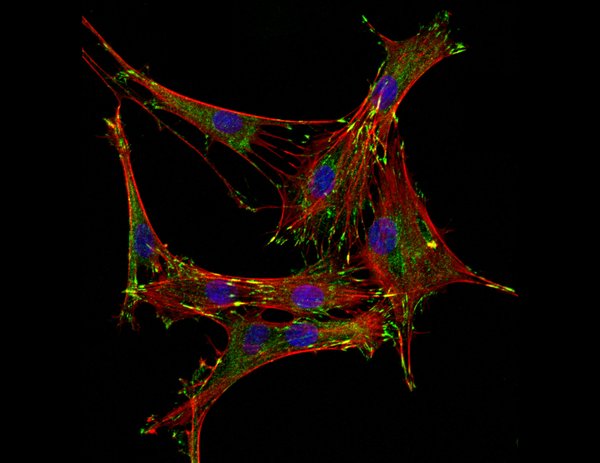
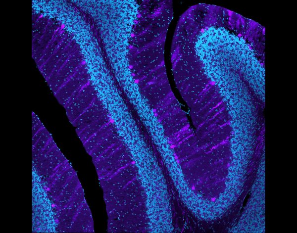
The Nikon A1R is an inverted, laser scanning confocal microscope with a stage-top Pathology Devices, Inc. environmental chamber.
This microscope uses point scanning laser illumination, and a single optical pinhole is placed within the emission path at the conjugate focal plane to reject out-of-focus light. As a result, this microscope captures light from an individual optical section. For this reason, the Nikon A1R is exceptional for volumetric imaging of cells under adherent and 3D environments. Multiple optical sections can also be captured and combined to generate a 3D image. Compared to the spinning disc confocal, the diameter of the pinhole can be adjusted to match the excitation wavelength and objective of the system, which allows for much more accurate 3D images of thick samples. This microscope is well-suited for applications requiring multi-dimensional imaging.