Bruker TruLive3D Light Sheet Microscope update
The Beckman Center Light Sheet Specialist is delighted to share some exciting news about our two specialized Light Sheet Microscopes.
The Bruker TruLive3D Light Sheet Microscope was purchased in 2023 by the Beckman Center and was first made available to targeted groups only. We have the pleasure to announce that it is now accessible to anyone on campus and beyond and is part of the ALMC booking system. Proceed as usual with our ticketing system to request a training on it. The Light-Sheet specialist will always be present with you during your imaging time to assist and help you with all your needs.
Several users recently got amazing results during multi-days, environment-controlled experiments. We will be show-casing their work on a portfolio page on our Wiki.
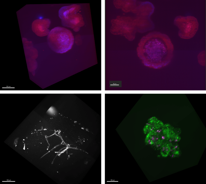
The Bruker TruLive3D is specifically designed to image organoids, spheroids, small embryos and single cells. Acquiring multiple z-stack is a breeze with this novel instrument and gives you direct 3D imaging. Each multi-days experiment will produce a time-lapse of your sample growing, ready to analyze with our Imaris workstation.

Single Objective Light Sheet Microscope
The second light sheet microscope of the Beckman Center is a Single Objective Light Sheet Microscope. This original, custom design microscope is ideal for single cells, small organoids or any first 1-3 layers of cells of a sample. The first prototype is now operational for a limited number of users but will gradually be made accessible for a wider audience so stay tuned!


Recent publications
- Guanhui Wu, Erin Taylor, Daniel T Youmans, Nausica Arnoult, and Thomas R Cech. Rapid dynamics allow the low-abundance RTEL1 helicase to promote telomere replication. Nucleic Acids Research 53, gkaf177 (2025). Microscope(s): Nikon NSTORM.